Trusted pipelines.
No code.
Easy install.

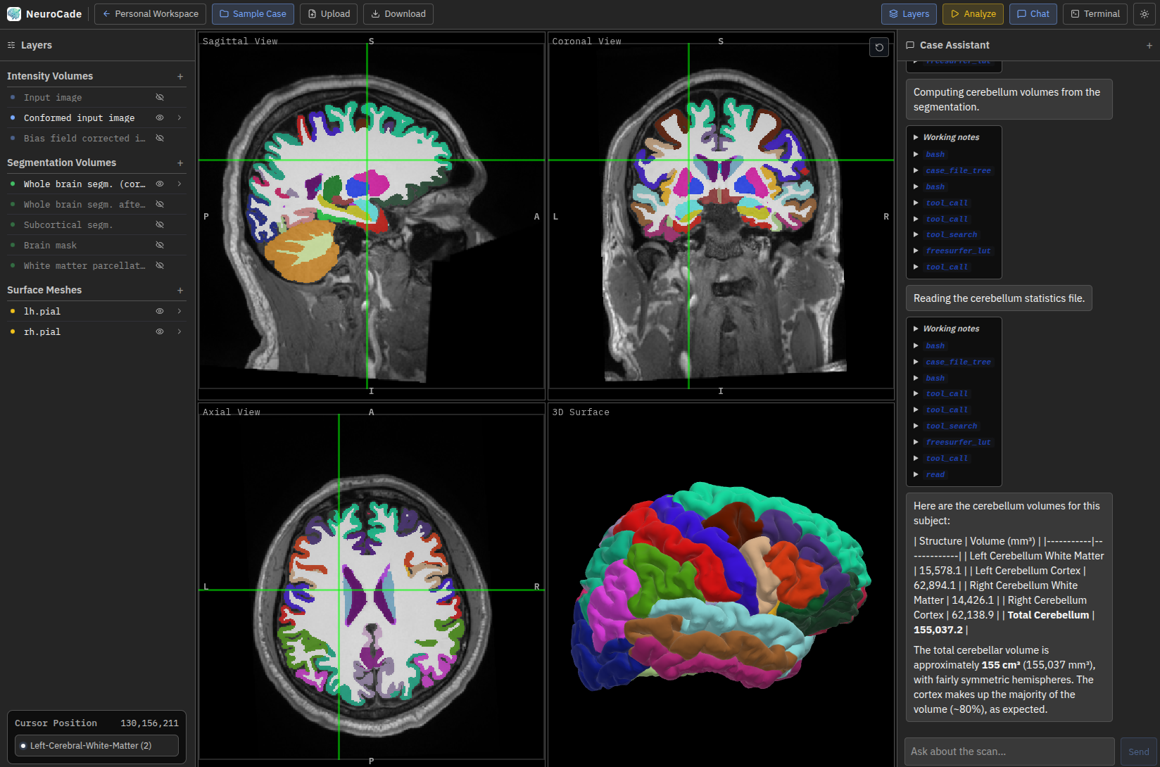
NeuroCade combines a familiar MRI viewer with built in support for FastSurfer and other establsihed NeuroImaging tools — letting you run complex neuroimaging workflows through the click of a button or AI chat.

Demo page
NeuroCade sample case workspace showing MRI views, layers, and assistant panel
AI Chat

Ask anything about
the scan in front of you

The AI assistant has direct context of the open study — slice, orientation, window settings, and patient metadata. No copy-paste, no switching tabs.

Real-time scan context

Every response is grounded in the active viewport — orientation, series, and slice position are automatically passed.

Structured report generation

Request a radiology report in plain English. NeuroCade produces structured DICOM SR-compatible summaries instantly.

Differential reasoning

Ask "what else could explain this?" and receive ranked differentials with supporting imaging criteria.

Assistant
Run hippocampal segmentation and compare to age-matched norms.
NeuroCade Running FreeSurfer subcortical segmentation on the loaded T1 MPRAGE…
Left hippocampus3.18 cm³
Right hippocampus3.41 cm³
Asymmetry index6.8% ↑
Age-norm percentile (62F)48th
Is the asymmetry significant?
NeuroCade An asymmetry index below 10% is generally within normal variation. At 6.8%, this is not considered significant. Left-right volumetric difference is not indicative of focal atrophy in this context. No additional workup recommended on volume grounds alone.
Ask a follow-up…
Neuroimaging Workflows

Complex pipelines, one sentence

Describe what you need in plain language. NeuroCade selects, configures, and runs the appropriate pipeline — no scripting required.

01 / DESCRIBE

Tell NeuroCade what you need

Speak naturally. No tool names, flags, or parameters required.

You said: "Segment all subcortical structures and flag anything below the 10th percentile for her age."
02 / PLAN

Pipeline is selected and explained

NeuroCade maps your intent to a validated pipeline and explains each step before running.

Plan: FSL FIRST → subcortical volumes → age-norm comparison (ADNI normative)
03 / RUN

Live progress in the viewer

Watch segmentation overlays appear in real time as the analysis completes.

Status: ▶ Running FSL FIRST
✓ Left thalamus
✓ Right thalamus…
04 / REVIEW

Structured findings, ready to sign

Results surface in-viewer with natural language interpretation, linked directly to the scan.

Finding: Left putamen 3.2 cm³ — 8th percentile. Recommend correlation with available history.
Analysis & Reporting

Findings that speak
your language

Quantitative results are always accompanied by contextual interpretation — not just numbers, but what they mean for this patient.

Subcortical Volumes — 64F

Age-normed
L. Hippocampus
3.18 cm³
R. Hippocampus
3.41 cm³
L. Amygdala
1.41 cm³
R. Amygdala
1.68 cm³
L. Thalamus
6.44 cm³
R. Thalamus
6.31 cm³
Total Brain Vol.
1,142 cm³

AI Interpretation

Auto-generated
Summary Global brain volume is within normal limits for age. Left amygdala volume falls below the 15th percentile — mild asymmetry noted. Hippocampal volumes are symmetric and age-appropriate. No evidence of medial temporal lobe atrophy pattern consistent with early AD.
Key Findings
✓ Hippocampi normal ⚠ L. amygdala asymmetry ✓ No cortical atrophy ✓ WM volume intact

Report Draft

Export ↗
"Volumetric MRI brain with subcortical segmentation performed. Hippocampal volumes are within normal limits bilaterally. Mild left amygdala volume reduction noted (1.41 cm³, ~15th percentile). No findings to suggest neurodegenerative atrophy pattern. Correlation with available history recommended."
Integrated Tools

The full neuroimaging
toolkit, voice-activated

NeuroCade wraps industry-standard neuroimaging tools in a conversational interface. No command line. No configuration files.

Segmentation

FreeSurfer & FSL

Subcortical segmentation, cortical parcellation, lesion mapping, and white matter analysis on any T1 or FLAIR series.

"Parcellate cortex and give me mean thickness per lobe."
Registration

Multi-modal Alignment

Automatic co-registration of T1, T2, FLAIR, DWI, and PET — aligned and overlaid without manual landmark placement.

"Overlay the FLAIR on the T1 and highlight any mismatch."
Diffusion

Tractography & DTI

FA, MD, RD, and AD maps generated from DWI. Probabilistic tractography on named white matter tracts.

"Show the corticospinal tract bilaterally."
Perfusion

ASL & DSC Maps

Cerebral blood flow, blood volume, and transit time maps with automated region-of-interest extraction.

"Compare CBF left vs right MCA territory."
Normative

Age-Matched Comparisons

Every volumetric result is automatically benchmarked against ADNI, HCP, and UK Biobank normative databases.

"Flag anything outside the 10th percentile for her age."
Reporting

Structured Reports

Findings are compiled into editable radiology reports, exportable as structured text, PDF, or DICOM SR.

"Draft a report for the referring neurologist."

Need help? Reach out!

Email